For Patients – Webinar 2020/09/22

Thank you for your attendance at our Webinar!

The webinar was attended by 44 participants.

Doctors answering the questions: Joshua Sonett (Thoracic and Cardiac Surgery), New York-Presbyterian/Columbia University Medical Center, New York, USA, Arun Rajan (Medical Oncology), National Cancer Institute, National Institutes of Health (NCI/NIH), Bethesda, USA, Edith Marom – President of ITMIG (Diagnostic Radiology), The Chaim Sheba Medical Center, Ramat Gan, Israel and Malgorzata Szolkowska (Pathology), National Tuberculosis and Lung Diseases Research Institute, Warsaw, Poland

Moderator: Pam Bruce, Executive Director of ITMIG

RECORDING OF THE WEBINAR IS NOW AVAILABLE

Q&A – Outstanding questions not answered during webinar

How to prevent recurrence and metastasis?

AR: The chances of recurrence and metastasis depend on the stage and subtype of disease at diagnosis. Early-stage disease should be treated aggressively with surgery (and post-operative radiation in select cases, especially in patients with thymic carcinoma) to decrease the chances of recurrence. Locally-advanced disease also merits aggressive multimodality management and these cases should ideally be discussed in a multidisciplinary tumor board setting to formulate an optimal plan of management. Complete surgical resection of early-stage or locally-advanced disease is one of the most important factors in reducing the risk of recurrence and metastasis in addition to tumor stage and the subtype of thymic tumor.


Is flu vaccine recommended?

AR: For most patients with no history of thymoma-associated autoimmune disease or treatment-induced autoimmunity, it should be okay to receive an inactivated influenza vaccine. However, for patients with a history of thymoma-associated autoimmunity or in the presence of an active autoimmune disease, or in patients who have previously experienced immune-related toxicity with immunotherapy, there is a risk of worsening of autoimmunity due to additional immune stimulation caused by the influenza vaccine and caution should be exercised in these cases.

In summary, the benefits of receiving an influenza vaccine are likely to outweigh the risks for most patients with thymoma who do not have an active or poorly controlled autoimmune condition.

On a related note, the Yellow Fever vaccine is contraindicated in patients with thymoma due to underlying defects in immune function.

Should a Covid -19 vaccine be available. Would you recommend taking it, considering that we are not suppose to take live vaccines?

AR: An excellent and very timely question. Various vaccine candidates are in evaluation for SARS-CoV-2 and the answer to this question ultimately depends on which vaccine candidate is approved for use and what the experience has been with that particular vaccine in clinical trials. It should be noted that in severe cases, Covid-19 causes a clinical picture which is very similar to that observed in patients with thymoma experiencing widespread immune activation either due to treatment (e.g., as a complication of immunotherapy) or due to the disease itself (thymoma-associated multi-organ autoimmunity).

Hence, upon approval of a safe and effective Covid-19 vaccine (preferably not a live vaccine), for most patients with thymoma who do not have a clinical history of autoimmunity or an active autoimmune condition, the benefits of vaccination against Covid-19 might outweigh potential risks (please also refer to our response for influenza vaccination below) and it might be reasonable to consider vaccination.

However, in the absence of an approved vaccine against Covid-19 at this time and uncertainties about what type of vaccine will be approved eventually, no recommendations can be made at present about the potential use of a Covid-19 vaccine in patients with thymic epithelial tumors.


Is immunotherapy safe if you have Myasthenia Gravis?

AR: Immunotherapy (of any kind) should NOT be used for treatment of thymoma in patients with a history of myasthenia gravis or other autoimmune conditions associated with thymoma. It can cause life-threatening complications.

On the other hand, immunosuppressive therapy used for treatment of myasthenia gravis is not the same as immunotherapy used for treatment of thymoma.

Is it safe to say that residual tumor on vena cava can be monitored with catscan with no contrast? I ve developed allergy to contrast.

EM: There is a solution for patients with an allergy to iodinated contrast, the contrast used with chest CT scans: performing an MRI or performing the contrast-enhanced chest CT after steroid preparation for the iodinated contrast. I would like to elaborate on the need for it so that we understand why contrast is needed when performing a CT.
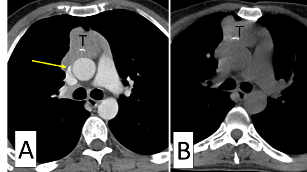
When imaging mediastinal tumors or residual tumors, which abut vessels, it is difficult to even impossible to distinguish the tumor itself from the vessel the tumor abuts, by CT. The contrast resolution of CT, which means its ability to distinguish the density of one soft tissue from the other, is low compared to, for example, MRI.  It is because of this that we inject contrast (iodinated contrast) at CT, to color the vessels (in white) so that we can assess whether the tumor invades the vessel, narrows the vessel, pushes the vessel, and also enables us to follow the tumor by measurements to plan treatment. Image A is a chest CT after iodinated contrast was injected. It shows the mediastinal tumor, a thymoma, marked with the letter T. The tumor abuts the mediastinal vessels and can be distinguished from them as they are colored by the injected contrast. We see that the superior vena cava is involved and narrowed in one location (yellow arrow pointing to it). This is information that is needed for planning surgery, planning radiation and even follow-up to assess if the tumor is shrinking or growing with chemotherapy. Image B, a chest CT without any contrast injection, is a chest CT performed the same day as image A, at a similar level. Notice, how one cannot even see the superior vena cava, nor tell whether it is involved or not. One cannot even tell if the other vessels are involved or measure this tumor’s entire circumference because it merges with the surrounding vessels, which we cannot tell apart.

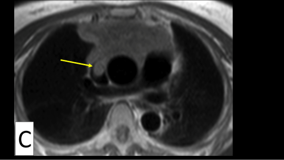
When a patient has a tumor or residual tumor that abuts a vessel, there are two ways in which one can overcome an allergy to iodinated contrast. One is to get premedicated with steroids prior to imaging with iodinated contrast. This is a good option if the allergy was not life threatening. Another option is to follow this tumor with MRI. The images on MRI are not as sharp as the images with CT, but MRI has superior contrast resolution. This means that MRI can better distinguish one soft tissue from the other, even without the use of contrast. Also, the intravenous contrast used at MRI, is not iodine based so it can be given safely when needed. The image below, image C, is an image of a different patient with thymoma. It is an MRI image performed without the use of any contrast. In this particular image, the method used is a ‘black blood method’ in which the vessels are black. One can clearly see, even though there is no intravenous contrast on board, that there is tumor invasion into the superior vena cava (which the yellow arrow points to).


There are more advantages and disadvantages to performing a CT as compared to MRI, all of which depend on each patient’s tumor and particular situation. Thus, the decision, on whether to follow with a chest CT and when to follow with MRI is a decision that should be made with your treating clinician. If the treating clinician is not sure, he/she could consult with his chest radiologist for further advice. This is why these tumors should be treated and followed in a medical facility that has a special interest in thymic tumors. A center in which all the subspecialties needed in the follow-up and treatment of this disease have a special interest in thymic tumors, including and not limited to thoracic surgery, medical oncology, radiotherapy, diagnostic radiology and neurology.

If the primary site (thymus) appears small and resectable, is there ever a place for removing the primary and then addressing residual disease/mets with systemic therapy?

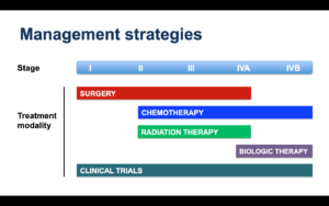
AR: Metastatic disease that is present outside the chest cavity (for example, involving the liver, bones, adrenal glands or other organs) should be treated with primary chemotherapy and surgery would generally not have a role in management. Removal of the primary site is unlikely to alter the course of disease in these situations and it is not recommended.

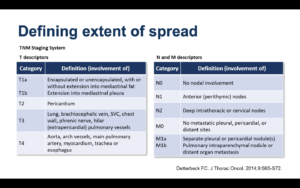
However, if there is limited metastatic involvement of the pleura or pericardium (stage IVA disease), there is a potential role for multimodality therapy with induction chemotherapy followed by surgery (with removal of the primary tumor and pleurectomy, pericardiectomy, or both) and radiation therapy, if indicated. It is highly recommended that these cases be discussed in a multidisciplinary tumor board setting in order to device an optimal strategy for management.


To what extent does pemetrexed affect spread of disease if it doesn’t eliminate it?

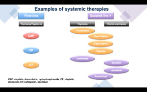
AR: Pemetrexed is active against advanced thymomas (much less so against thymic carcinomas). It can shrink growing tumors and is effective in preventing the spread of disease and development of new lesions. Most patients tolerate treatment very well and treatment is often associated with durable response and disease control (from many months to several years).


Where can I find copies of all research papers that have to do with treatment efficacy and prognosis of stage IVB squamous thymic carcinoma cancer?

MSz: PubMed database is one of the most popular sources for research papers. Click the link:  https://pubmed.ncbi.nlm.nih.gov and use the filter e.g. “treatment IVB thymic squamous cell carcinoma”.

What’s your view if only immunotherapy (no surgery) has been used for stage 4A with lung pleural effusion and nodules? After 40 infusions, lungs appear clear with CT scan.

AR: Use of immunotherapy for previously untreated thymic cancers (i.e., no prior chemotherapy) would not be considered standard therapy.

If immunotherapy has been used for a period of two years or more in this setting and has been associated with a very good partial response or a complete response, it would be reasonable to stop treatment and continue with active radiological surveillance with CT scans approximately every three months. Treatment can be resumed if there is evidence of disease activity on follow-up scans. This paradigm of stopping immunotherapy after two years in case of a very good response and restarting it if there is evidence of disease activity, is increasingly used for treatment of lung cancer.


In regards to pregnancy and thymoma, Dr. Rajan spoke of non active disease and it being safe; however, I wondered about having active disease, not currently in treatment and whether it is ok to get pregnant although with the uncertainty of future scans? I have had some recent growth in a nodule within the past month as I was working with a fertility specialist.

AR: First, chemotherapy and other systemic anti-cancer therapies should be avoided during pregnancy, especially during the first 12-14 weeks, which is a critical period for fetal development. In situations where initiation of anti-cancer therapy is absolutely unavoidable, treatment is delayed until after the first trimester.

In this particular situation, if thymoma has been generally stable for months or years, the chances of significant growth within a period of a few months is low (especially, if histology is WHO subtype A, AB, B1 or B2). However, this is not guaranteed since the rate of growth can change over time. Therefore, although it is theoretically possible to have an uneventful pregnancy in the presence of quiescent disease, there is a risk that the rate of growth might increase during the course of pregnancy and additional treatment might become necessary.

If there is only one nodule growing within the past month and all other sites have a history of stability over a long period, one option is to consider local therapy for the growing nodule to treat it adequately and achieve overall disease stability before pregnancy.

For treatment of a solitary site of site of growth during pregnancy, surgery would carry with it risks of exposure to anesthetic medications. Radiation therapy should be avoided. Another option is thermal ablation (cryoablation or microwave ablation), which might be relatively safe under these circumstances and has been used to successfully treat solitary sites of active thymoma.

Ultimately, this is a complex question and requires a detailed discussion between a patient, her obstetrician and the oncology team.
ITMIG